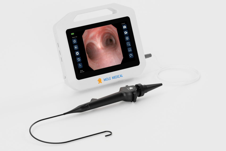
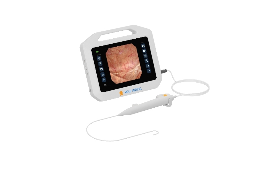
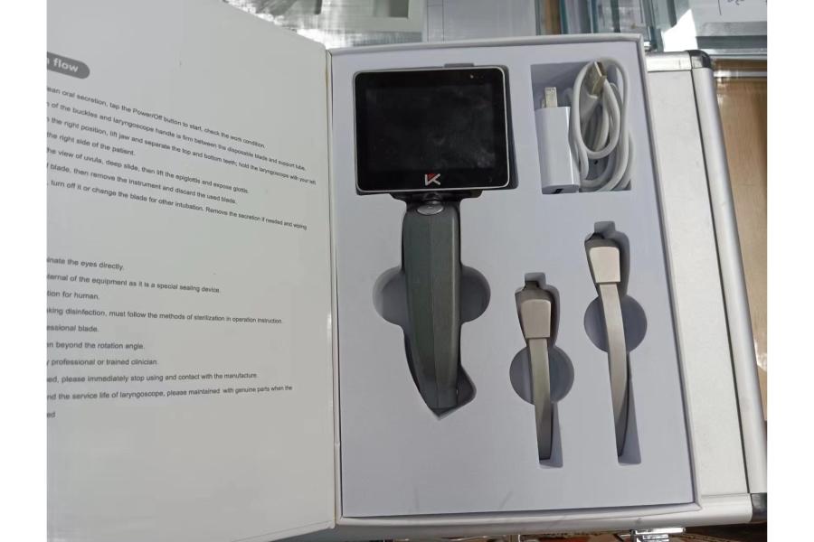
How to use anesthesia laryngoscopy?
Sep 04, 2023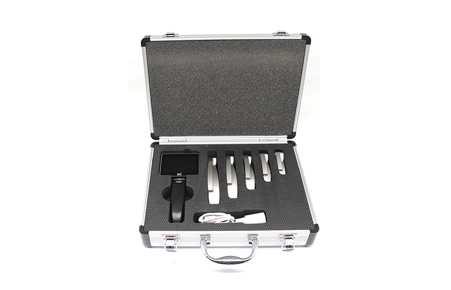
1.Direct anesthesia laryngoscopy is performed under mucosal surface anesthesia. The surgeon holds the mirror in his left hand and protects the upper teeth with thick gauze…
Anesthesia laryngoscopy
1.Direct laryngoscopy is performed under mucosal surface anesthesia. The surgeon holds the mirror in his left hand and protects the upper teeth with thick gauze. The mirror is introduced into the mouth along the back of the tongue and transferred deep into the midline, to the base of the tongue. The thumb and index finger of the right hand assist in holding the tube from front to back when the epiglottis is seen from the laryngoscope. Tilt the proximal end of the laryngoscope up (tilted forward when sitting) and the distal end toward the back of the pharynx without touching it. Continue into the lens beyond the free edge of the epiglottis. After seeing the epiglottis tubercle, lift the laryngoscope with the left hand with a parallel upward force, and press the epiglottis to fully lift it to expose the larynx. At this time, if laryngospasm occurs, the glottis is closed and the glottis cannot be seen.
2.The laryngoscope should be fixed in place, and the laryngeal image can be seen after the laryngospasm touches for a moment. If the laryngoscope touches the mucosa of the laryngeal cavity too deeply to cause spasm of the arch reflex, the laryngoscope should be slightly retracted for observation, and the subject should be told to make the sound of “clothes” and observe the movement of the vocal cords. At this time, the surgeon can perform various necessary operations with the right hand.
3.If the subject’s neck is short and thick, and the anterior commissure of the vocal cords is not easily exposed, the head must be raised, the left hand lifts the laryngoscope up, the right thumb presses the laryngoscope up from the bottom, and the remaining fingers of the right hand should be fastened on the patient’s right upper The teeth are supported in unison to lift the epiglottis. If this approach is unsuccessful, you can ask your assistant to compress the thyroid cartilage down or use a combined laryngoscopy instead. An anterior arthrolaryngoscope not only provides a clear view of the anterior vocal fold joints, but can also be inserted into the glottic fissure to examine the subglottic space.
4.When examining young children, in order to prevent laryngeal edema after this operation, the tip of the laryngoscope should not compress the epiglottis, and the base of the tongue should only be lifted forward, and the epiglottis will stand up to expose the larynx.
5.Complications of anesthesia laryngoscopy
It usually happens rarely. Children, especially those with spasticity, may develop severe, life-threatening laryngospasm during surgery. During the operation, the movements should be as gentle as possible to reduce the damage to the throat mucosa and reduce the chance of hematoma, bleeding or bleeding secondary to infection.


Latest Articles
Clinical comparison of foreign body removal procedures using rigid bronchoscopy, fiberoptic bronchoscopy, and flexible electronic bronchoscopy
Bronchial foreign bodies are a common emergency in pediatrics. Clinically, bronchoscopy techniques are typically used to remove the foreign bodies. Currently, the three main bronchoscopy techniques each have their own characteristics, and among them, the flexible bronchoscopy shows unique clinical value in pediatric patients. This article conducts a clinical application analysis of all three bronchoscopy ... Read more

How Fibre Optic Laryngoscopes Improve ENT Procedures
In modern ENT procedures, precision and visibility are key. That’s where the laryngoscope fibre optic technology comes in. Unlike traditional tools, these advanced devices use fibre optics to provide a clear, well-lit view of the throat and vocal cords. This means doctors can see more and do more—with less risk to the patient. But how ... Read more
Flexible Laryngoscopy: A Clearer Voice for Quicker Diagnoses
Flexible Laryngoscopy is a powerful tool that helps ENT specialists do just that. It uses a thin, flexible scope to view the throat, vocal cords, and airway in real-time. The procedure is quick, non-surgical, and performed right in the clinic. For patients with voice changes, chronic cough, or throat discomfort, this method offers fast answers ... Read more
Smart Solutions in Ureteroscopy: Mole Medical’s Disposable Flexible Scope vs Karl Storz Legacy
In the field of urology, the name “ureteroscope Karl Storz” has long stood for precision and reliability. As a global leader, Karl Storz has set the standard for reusable ureteroscopes. But now, a new contender is entering the spotlight. Jiangsu Mole Medical, a national high-tech enterprise, is challenging the status quo. With a focus on ... Read more
Behind the Scenes with Mole Medical: Inside the World of Leading Video Laryngoscope Manufacturers
In the fast-evolving medical device industry, video laryngoscope manufacturers play a key role in advancing patient care. Mole Medical stands out not just for its products, but for what happens behind the scenes. What makes this company different? Why do so many professionals trust its solutions? One reason is speed. Mole Medical has an R&D ... Read more