Double Lumen Endotracheal Tube Placement Guide
Dec 23, 2023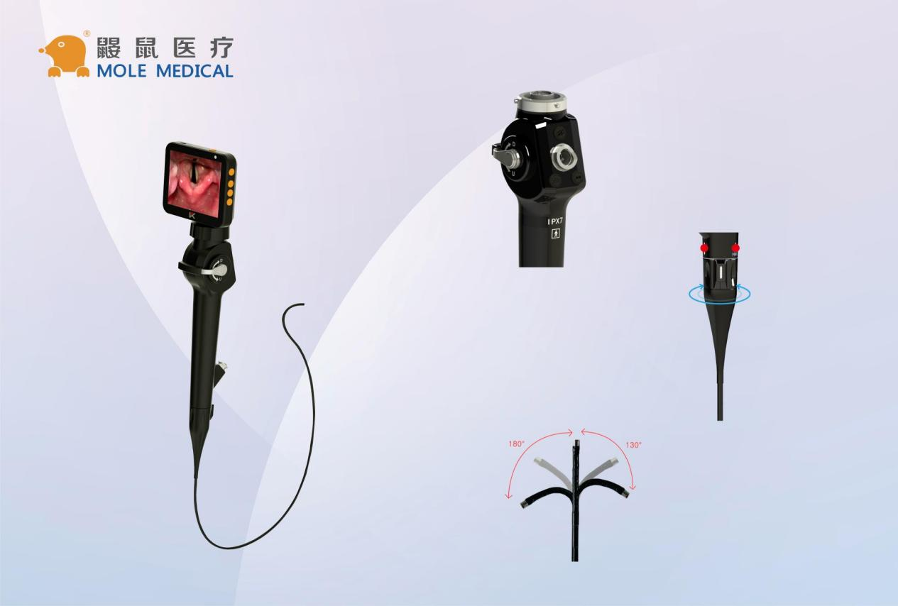
Double lumen endotracheal tube placement is a crucial element of thoracic surgery. Its successful execution ensures lung isolation, which is vital for complex surgical procedures in the chest cavity. This guide provides a comprehensive overview of double lumen endotracheal tube placement, covering its design, advantages, and role in anesthesia. Various insertion techniques, confirmation methods, and airway management strategies during thoracic surgery are also discussed.
Understanding the procedure and following best practices ensures a successful outcome. Through careful consideration of the discussed factors, optimal double lumen endotracheal tube placement can be achieved, further contributing to better health outcomes for patients undergoing thoracic surgery.
This comprehensive guide emphasizes the importance of considering every aspect of double lumen endotracheal tube placement, highlighting the role of medical professionals in providing quality care. By following the recommended practices, healthcare practitioners can ensure successful procedure and improved patient outcomes.
Read on to learn more about double lumen endotracheal tube placement, its significance in thoracic surgery and the necessary steps for optimal lung isolation.
Keywords: double lumen endotracheal tube placement, thoracic surgery, lung isolation
Understanding Double Lumen Endotracheal Tubes
Double lumen endotracheal tubes (DLT) are specialized devices designed to provide lung isolation during thoracic surgery. They consist of two tubes, one for the trachea and the other for the bronchus leading to the lung to be isolated. This design allows for independent ventilation of each lung, making it possible to operate on one lung while controlling the airflow in the other.
The advantages of DLT over other methods of lung isolation include greater control over ventilation and the ability to perform one-lung ventilation. One-lung ventilation, wherein only one lung is ventilated while the other is collapsed, is a critical component of many thoracic surgical procedures.
DLTs are commonly used in surgeries such as lobectomy, thoracotomy, pneumonectomy, and esophagectomy. They are also useful in managing patients with collapsed lungs or airway injuries.
The selection of the appropriate size and type of DLT depends on multiple factors, including the patient’s medical history, weight, and height, as well as the type of surgery being performed. An anesthesiologist with expertise in airway management and intubation technique should be involved in the decision-making process.
Proper placement of a DLT is essential for effective lung isolation. In the next section, we will discuss the indications and considerations for DLT placement.
Indications and Considerations for Double Lumen Tube Placement
Double lumen endotracheal tubes are used in several situations to achieve effective one-lung ventilation in thoracic surgery. These tubes are commonly used for procedures such as bronchopleural fistula repair, lobectomy, and esophagectomy.
Anesthesiologists use double lumen tubes to achieve sufficient control over the patient’s airway while ensuring complete isolation of one lung. This is essential in thoracic surgeries where the surgeon needs an unobstructed view of the surgical site.
Before placement, there are critical considerations that must be taken into account. An anesthesiologist will start by assessing the patient’s airway before selecting the appropriate size and length of the tube. They will also consider the patient’s positioning, the type of surgery involved, and the necessary equipment to use.
A potential complication that an anesthesiologist faces during tube placement includes a possible tear or perforation of the trachea. This stems from the rigidity of the tube, which could cause undue stress on the airway.
When performed correctly, the use of a double lumen tube offers a dependable way of ensuring one-lung ventilation and lung isolation during thoracic surgical procedures. Its use requires a trained anesthesiologist to determine indications for use and the appropriate precautions for effective patient care.
Preparing for Double Lumen Tube Placement
Before placing a double lumen endotracheal tube, proper preparation is key to ensure a successful procedure. Anesthesia providers should carefully review the patient’s medical history and physical exam, paying special attention to any previous airway or lung pathologies.
The next step is to select appropriate equipment, including a fiberoptic bronchoscope, and confirm its proper operation. The operating room should be equipped with all necessary airway equipment, such as suction, an oxygen source, and rescue devices.
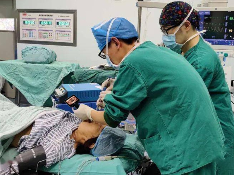
Patient positioning is critical for proper placement of the double lumen endotracheal tube. For thoracic surgery, the patient should be positioned on their side, with the operative side tilted upwards to facilitate lung isolation. The anesthesia team should carefully position the patient to prevent pressure points and other complications.
Proper communication and teamwork between the anesthesia provider and the surgical team is essential to ensure a smooth procedure. Clear communication regarding the timing of the procedure, anticipated anesthesia duration, and any special instructions will help prevent complications and ensure a successful outcome.
Techniques for Double Lumen Tube Insertion
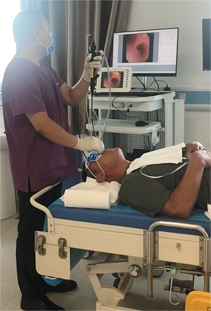
Inserting a double lumen endotracheal tube is a critical step in achieving effective lung isolation during thoracic surgery. There are various techniques for performing this procedure, including direct visualization and the use of fiber-optic bronchoscopy.
Direct visualization involves using a laryngoscope and placing the double lumen tube under direct vision. This technique is useful when the anatomy of the airway is easily identifiable and the patient’s neck is adequately extended.
The use of fiber-optic bronchoscopy allows for greater visualization of the airway anatomy and can be especially helpful when the patient’s airway is difficult to manage. Additionally, this technique can be used to confirm proper placement of the double lumen tube.
It is essential to evaluate the patient’s individual needs and anatomical considerations when determining which technique to use. Choosing the appropriate insertion technique can significantly impact the success of the procedure and the patient’s outcome.
Regardless of the technique used, ensuring proper placement and positioning of the double lumen tube is crucial to achieving effective lung isolation during thoracic surgery.
Confirming Placement and Troubleshooting
Ensuring the correct placement of a double lumen endotracheal tube is crucial for effective lung isolation during thoracic surgery. Several methods can confirm the tube’s position, including auscultation, capnography, and fiber-optic bronchoscopy. Auscultation involves listening to breath sounds in both lungs to ensure equal ventilation. Capnography measures the carbon dioxide levels in exhaled air, with a difference in values indicating malposition of the tube. Fiber-optic bronchoscopy provides a direct visualization of the tube’s location and can also identify potential complications.
In case of malposition or complications, several troubleshooting strategies can address the issue. For instance, repositioning the patient, adjusting the tube’s depth, or inflating the bronchial blocker can help manage ventilation and achieve proper lung isolation. In rare cases of severe complications or tube dislodgment, transitioning to one-lung ventilation with a bronchial blocker may be necessary.
Overall, confirming the correct placement of a double lumen endotracheal tube and troubleshooting any issues are critical steps to ensure successful thoracic surgery with effective lung isolation.
Managing the Airway during Thoracic Surgery
Proper airway management is crucial during thoracic surgery after ensuring the double lumen endotracheal tube placement. The anesthesiologist must continuously monitor the patient’s oxygen saturation, end-tidal carbon dioxide, and blood pressure. They should also periodically evaluate lung function and adjust ventilator settings to maintain optimal lung isolation.
When performing one-lung ventilation, the ventilator settings may need to be adjusted to ensure that the non-ventilated lung does not collapse. The anesthesiologist may also need to administer additional medications to prevent coughing or bronchospasm.
In addition to these measures, the surgeon may use other techniques to minimize the risk of lung injury, such as applying positive pressure to the collapsed lung to prevent atelectasis.
Throughout the procedure, communication between the anesthesiologist and the surgeon is essential for timely adjustments to the patient’s ventilation and positioning. By working together, the medical team can ensure optimal outcomes for the patient.
Conclusion
Double lumen endotracheal tube placement is a critical component of effective lung isolation during thoracic surgery. By following the techniques and considerations outlined in this guide, anesthesiologists can ensure precise placement and management of the airway throughout the procedure.
Proper preparation, including patient positioning and equipment selection, is essential for successful double lumen endotracheal tube placement. Anesthesiologists should also be familiar with various techniques for insertion, including direct visualization and fiber-optic bronchoscopy.
Confirming the correct placement of the double lumen endotracheal tube is crucial and can be achieved through various methods, such as auscultation and capnography. In case of malposition or complications, quick troubleshooting strategies can be implemented to minimize negative outcomes.
Throughout the surgery, monitoring and maintaining lung isolation are paramount. Anesthesiologists should be vigilant of any factors that may compromise the airway, including patient positioning and hemodynamic changes.
By adhering to best practices for double lumen endotracheal tube placement, anesthesiologists can contribute to the success of thoracic surgeries and ultimately improve patient outcomes.


Categories
Latest Articles
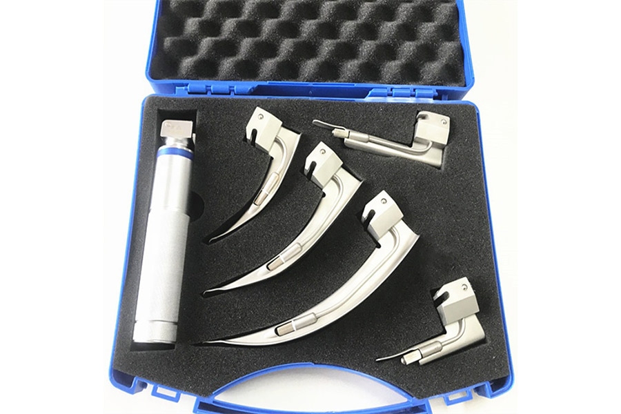
How Much Does the C-MAC Laryngoscope Really Cost?
C-MAC laryngoscopes work well. But they are expensive. The price depends on the model. It also changes with added features or tools. Some setups cost much more than others. Many hospitals do not have extra money. So, they look for other options. They want tools that still work but cost less. This leads to a key question. ... Read more
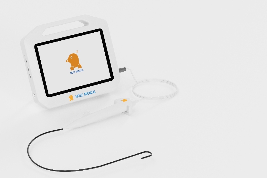
Mole Medical: Leading Supplier of Advanced Video Laryngoscopes for Safer Airway Management
Why is Precise and Safe Intubation Important? Intubation helps patients breathe when they cannot do it on their own. A clear airway is needed for surgery and emergency care. Doctors must place the tube quickly and correctly. Mistakes can cause injuries or delays. A precise and safe method is important for better patient outcomes. How ... Read more
Flexible Digital Disposable Ureteroscopes for Cost-Effective Solutions
How is Technology Changing Urological Procedures? Medical tools are improving fast. Ureteroscopy is now more precise and safer. New devices help doctors see inside the urinary tract clearly. Better imaging and flexible designs make procedures easier. Patients experience less pain and faster recovery. Hospitals need tools that improve accuracy and reduce risks. Why Choose a ... Read more
Affordable Bronchoscope: Price, Features, and What to Expect
The Role of Bronchoscopes in Modern Medicine A bronchoscope is an important medical tool. Doctors use it to look inside the lungs and airways. It helps diagnose infections, tumors, and other lung problems. It is also used to remove blockages and take tissue samples. Hospitals and clinics rely on bronchoscopes for many procedures. A clear ... Read more
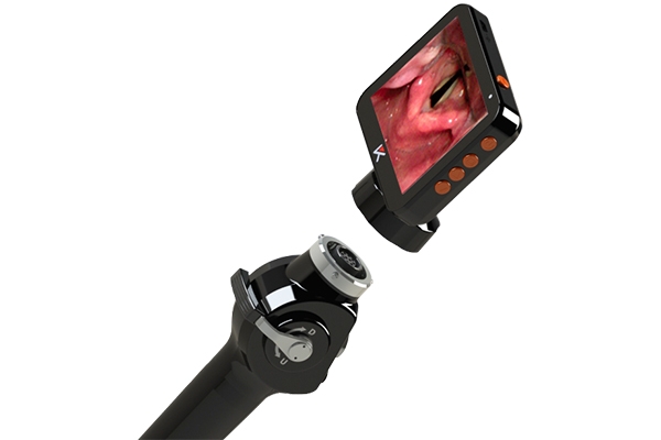
Laryngoscope mole Fiber optic: A Clearer View, A Safer Airway
The Laryngoscope mole Fiber optic is a tool for airway management. It helps doctors see inside the throat. It has a fiber optic system that gives a bright and clear view. Doctors use it to guide breathing tubes into the airway. It makes intubation easier and safer. This laryngoscope works with different blades. The Miller blade ... Read more